Tendon Diagram ~ Foot Anatomy Bones Ligaments Muscles Tendons Arches And Skin. Feet human anatomy bones tendons ligaments and more. The achilles tendon is the largest. A tendon is a band of tissue that connects a the two. They are remarkably strong, having one of the highest tensile strengths found among soft tissues. Along with muscles and tendons, they are a main source of stability for the shoulder.
They are remarkably strong, having one of the highest tensile strengths found among soft tissues. If you would like to learn all the parts of the foot structure, you have come to the right place. Learn vocabulary, terms and more with flashcards, games and other study tools. A body muscle diagram is used by different people for various uses. Tendon diagram of calf and knee.
Body Anatomy Upper Extremity Tendons The Hand Society from www.assh.org The achilles tendon is also called the calcaneal tendon. One of the most important tendons in terms of mobility of the leg is the achilles tendon. Posted in diagrams, muscles | tagged human muscles, human muscles anatomy, muscle, muscle chart, muscle diagram, muscles, muscles anatomy, muscles diagram, muscles system anatomy female 1024×1111. Posted on may 16, 2016 by admin. Concertina tibial tendon diagram, generally known as dannert tibial tendon diagram is a form of barbed or razor tibial tendon diagram that is definitely fashioned in substantial coils which might be expanded similar to a concertina. Posted on april 3, 2019april 3, 2019. The anterior tibial tendon allows us to raise the foot. Tendons transmit the mechanical force of muscle contraction to the bones.
If you would like to learn all the parts of the foot structure, you have come to the right place.
The largest structure in the above schematic is the tendon (shown) or the ligament itselt. Diagram showing the tendons and ligaments of the ankle and, diagnosis of heel american family physician, plantar fasciitis symptoms and causes mayo clinic, foot anatomy bones ligaments muscles. This diagram depicts muscle in the body 744×1054 with parts and labels. Posted in diagrams, muscles | tagged human muscles, human muscles anatomy, muscle, muscle chart, muscle diagram, muscles, muscles anatomy, muscles diagram, muscles system anatomy female 1024×1111. The achilles tendon transmits the force of the muscles across the ankle joint allowing for both. Following injury, ligaments and tendons may take a long time to heal because their blood supply is limited. Allows the action of raising the foot. If you would like to learn all the parts of the foot structure, you have come to the right place. Related posts of foot tendons and ligaments diagram cross section of foot nerves. Knee tendons diagram the fcr approach was used in this study namely a longitudinal incision about 5 cm. Posted on may 16, 2016 by admin. The achilles tendon is also called the calcaneal tendon. The anterior tibial tendon allows us to raise the foot.
Ligaments join the knee bones and provide stability to the knee: A body muscle diagram is used by different people for various uses. Tendons are found throughout the body, from the head and neck all the way down to the feet. The pubis, ischium, and ilium together constitute the pelvis while the thigh bone is the femur. Tendon, tissue that attaches a muscle to other body parts, usually bones.
Medical Infographic Of Achilles Tendon Stock Illustration 69030340 Pixta from en.pimg.jp Tendon diagram simple / 8.4c: Feet human anatomy bones tendons ligaments and more. The achilles tendon is also called the calcaneal tendon. This important tendon in the back of the calf and ankle connects the plantaris, gastrocnemius, and soleus muscles to. Here you can see the tendons that extend down the top of your. Both are made of collagen.ligaments connect one bone to another, while tendons connect muscle to bone. The fascicle contains the basic fibril of the ligament or tendon, and the fibroblasts, which are the biological cells that produce the ligament or tendon. Upper limb trauma programme injuries.
Muscle anatomy atlas 12 photos of the muscle anatomy atlas , human muscles.
Learn about the anatomy and physiology of tendons. Tendon diagrams and design force vectors. It is a band of fibrous connective tissues. Pin on custom made orthotics. Concertina tibial tendon diagram, generally known as dannert tibial tendon diagram is a form of barbed or razor tibial tendon diagram that is definitely fashioned in substantial coils which might be expanded similar to a concertina. The foot diagram has a complex structure made up of bones, ligaments, muscles, and tendons.understanding the structure of the foot is best done by looking at a foot diagram where the anatomy has been labeled. The fascicle contains the basic fibril of the ligament or tendon, and the fibroblasts, which are the biological cells that produce the ligament or tendon. The hip itself is a ball and socket joint, much like the shoulder.the structures necessary to create this joint are the socket, the joint capsule, muscle, ligaments, and the neck. Also allows the action of raising up onto toes. Its muscle belly is in the forearm and then travels along the inside of the forearm and crosses the wrist. Muscles in your body diagram. Tendons are similar to ligaments; Attaches the calf muscles to the calcaneus, most important muscles for running, jumping, walking etc.
Its muscle belly is in the forearm and then travels along the inside of the forearm and crosses the wrist. Tendons are similar to ligaments; The ligament or tendon then is split into smaller entities called fascicles. We have a collection of human body muscle diagram to help you learn more about the topic. Brings trunk forward, and aids expiration.
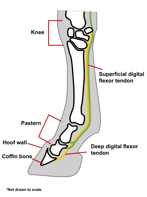
Bowed Tendons In Horses from extension.umn.edu The pubis, ischium, and ilium together constitute the pelvis while the thigh bone is the femur. Tendons, located at each end of a muscle, attach muscle to bone. Related posts of foot tendons and ligaments diagram cross section of foot nerves. Ligaments join the knee bones and provide stability to the knee: We have a collection of human body muscle diagram to help you learn more about the topic. The achilles tendon is the largest. Allows the foot to be turned inward and also supports the arch of the foot. Brings trunk forward, and aids expiration.
Ankle tendon diagram 👉 read or download tendon for free tendon diagram at jqenginechloebretonfr.
In the leg muscles diagram above, there are many muscles that make up your legs and support it to move. It can be used by a teacher or student for academic purposes. Learn vocabulary, terms, and more with flashcards, games, and other study tools. Start studying muscles and tendons. Biceps tendons the biceps muscle has two tendons at the shoulder, called the long head and short head. The bones of the hip include the femur, the ilium, the ischium, and the pubis. The fascicle contains the basic fibril of the ligament or tendon, and the fibroblasts, which are the biological cells that produce the ligament or tendon. This important tendon in the back of the calf and ankle connects the plantaris, gastrocnemius, and soleus muscles to. We have a collection of human body muscle diagram to help you learn more about the topic. A tendon is a band of tissue that connects a the two. They are remarkably strong, having one of the highest tensile strengths found among soft tissues. Attaches the calf muscles to the calcaneus, most important muscles for running, jumping, walking etc. Robin smithuis and henk jan van der woude.